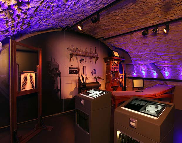
Roentgen Museum & Home, Germany

The picture below, is of the Roentgen Home and Museum.

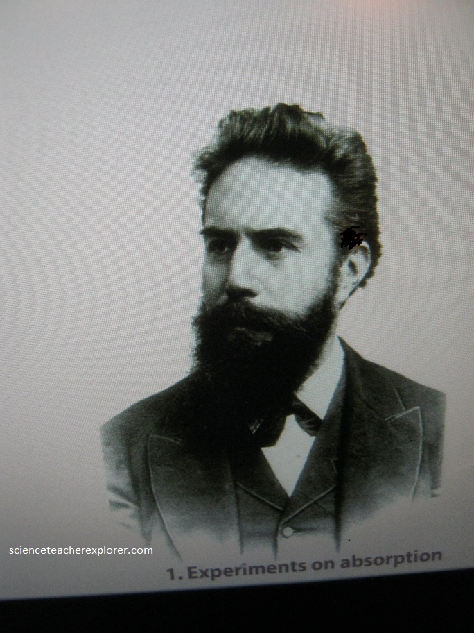
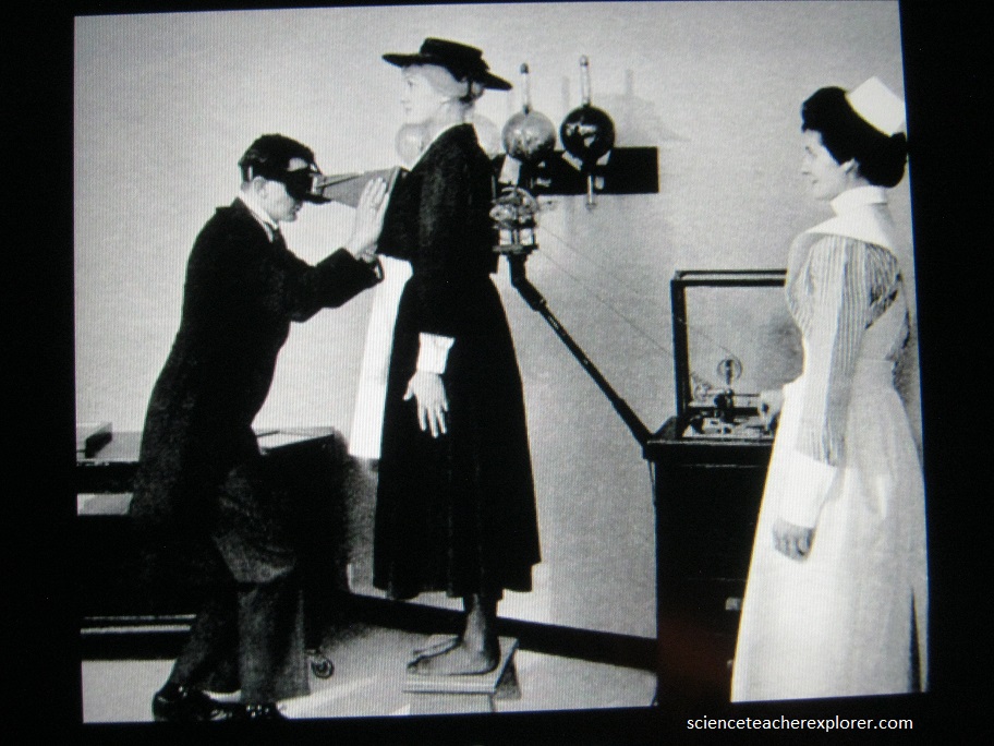
On August 8th, 2017; I took the day to explore this museum and study x-rays. The man who discovered x-rays was awarded the first ever Nobel prize in 1901. Wilhelm Conrad Röntgen was born in the Remscheid suburb of Lennep in 1845 and he has given his name, not only to the museum but – in German – to x-rays themselves.
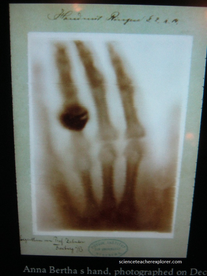
For the Germans, “to x-ray” is “röntgen”. Nowadays everything in the Deutsches Röntgen Museum in Lennep is connected with the invention which made Röntgen famous throughout the world: x-rays. Below is a historical image of the first X-Ray taken of the hand from Rontgen’s future wife.
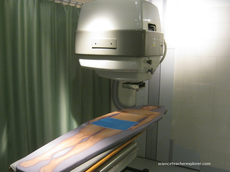
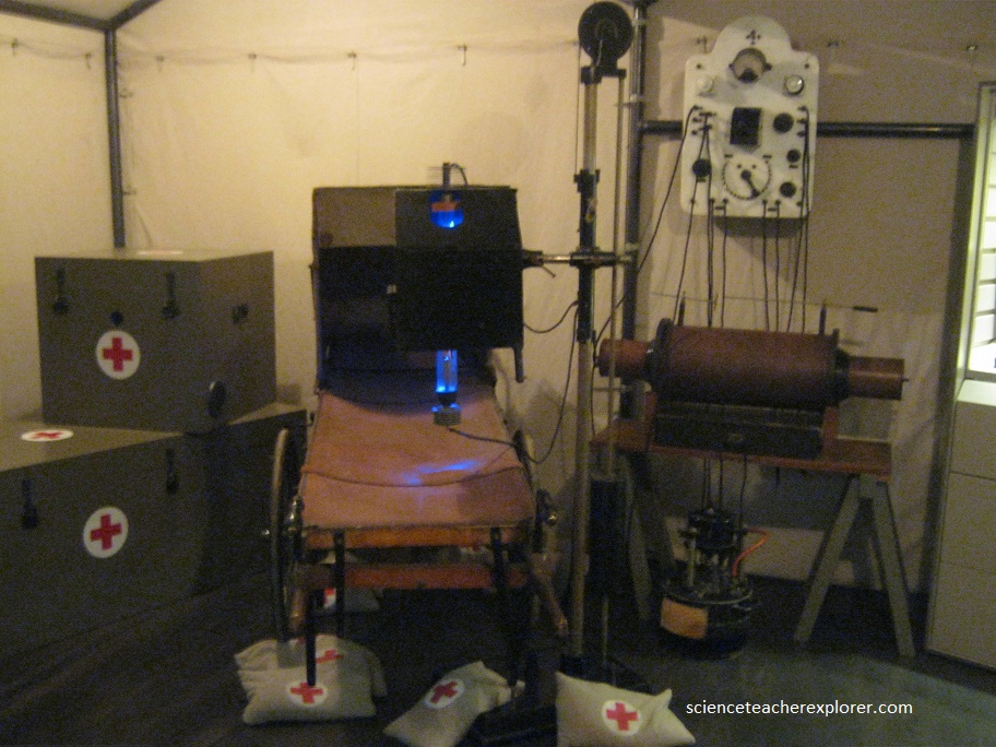
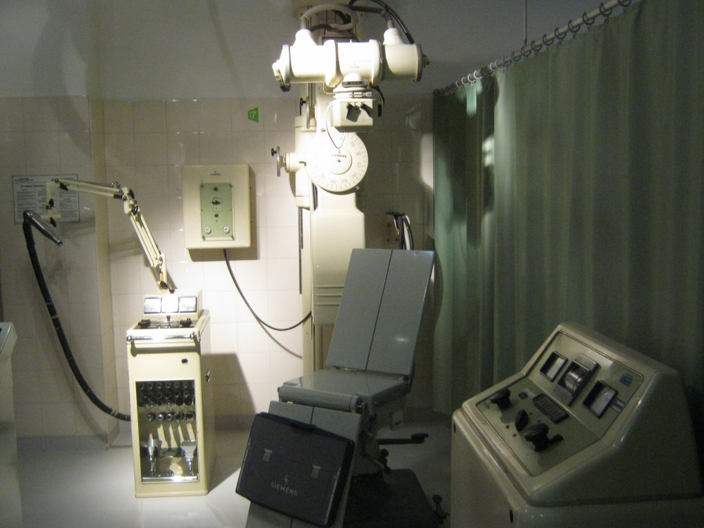
The range of X-Ray applications is countless. In the medical area alone, the museum had an impressive amount of apparatus ranging from the seemingly primitive “Diaphor R” x-ray instruments to a modern experimental machine for short-time tomosynthesis. Here it is possible to check things like wheeled rims on cars or survey welding points in pipelines. Paintings and art in general often require the accurate insights provided by x-rays, whether it be to illuminate a Rembrandt painting or a 900-year old Peruvian mummy. While visiting the museum, I traveled effortlessly through space and time, past and present of the x-ray world.
An X-ray machine is basically a camera, but it creates it’s own “light” in the form of X-rays. An electric current is sent to the vacuum tube that houses a ‘cathode’ at one end and an ‘anode’ at the other. The cathode is a filament, like in lightbulbs. When the current passes through it, it heats up and emits electrons, (negatively charged atomic particles), into a vacuum tube. The anode is positively charged, so it acts as a magnet and pulls those negatively charged electrons toward it. Embedded in the anode is a metal disk, usually made of tungsten. When one of those incoming electrons collides with a tungsten atom, that atom loses one of it’s own electrons, (so another electron in the atom jumps in to fill it).
The electron that jumps in comes from farther away from the atom’s nucleus, (and has much more energy than the one that got knocked out). When it fills in the empty spot, it has to release it’s surplus energy, (which has much more energy than the one that got knocked out). When it fills in the empty spot, it has to release its surplus energy, (which it releases as a ‘photon’). This happens billions of times in one X-ray procedure.
A lead container surrounds the vacuum tube to absorb X-rays. A small opening in the container allows the escape of photons, which first pass through a series of filters that stop all but a pure beam of X-ray photons.
Now we take the picture. On one side of whatever is being X-rayed, (say a human hand), is the window through which the beam is emitted; on the other side of the hand is the film. As the X-ray photons meet the atoms in the hand, they will either pass through or be absorbed by them. The softer tissues in the human body, (skin, organs and muscles), are made up of relatively small atoms. The X-ray photons pass right through them and leave their mark as the lighter areas on the film, just like light hitting a camera’s film. The atoms in the bones, (primarily calcium), are much larger, and they absorb the X-rays and stop them from reaching the film. Result: The film has captured an image of the bones in the hand.